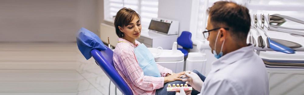
What is a Dental PPO?
Finding a dentist can be a cumbersome process for anyone. For those who don’t understand how their insurance works from a coverage standpoint, it can lead one to avoid making the appointment altogether...

Dental cone beam computed tomography (CT) is a quite special x-ray machine used in situations where regular facial or regular dental x-rays are not enough. It is not used widely because the radiation exposure from this scanner is little more than regular dental x-rays. This CT scanner uses a state-of-the-art technology to deliver three dimensional images, soft tissues, dental structures, bone and nerve paths in the craniofacial region in a single scan. There can be more precise treatment planning if the images are obtained with cone beam CT.
Dental cone beam CT might be used to produce images that are similar to those produced by conventional CT imaging. In case of cone beam CT, an x-ray beam in the shape of a cone is moved around the person to deliver a large number of images, also called views. Cone beam CT delivers precise images of the bone and is performed to evaluate diseases of the dentition, jaw, bony structures of the face, sinuses and nasal cavity. It does not provide the full diagnostic information in terms of soft tissue structures such as lymph nodes, muscles, glands and nerves.
A cone beam CT examination needs no special preparation. Before going through the exam, one may be asked to remove anything that may disrupt the imaging, including metal objects, such as eyeglasses, jewellery, hairpins and hearing aids. Dentist might also ask to remove the removable dental work but it is important to bring these to examination, as the dentist or oral surgeon may need to examine these as well. Women should always inform their dentist if they are pregnant.
Patient will be asked to stand or sit in the Radiology room. The dental assistant will position the patient according to the area of interest, centered in the beam. One will be asked to remain still while the x-ray source and detector revolve around for a 360-degree rotation or less. This usually takes about 15-20 seconds in which the entire mouth and dental structures are imaged, and it takes less than 10 seconds for a regional scan that focuses on a specific area of the maxilla.
The physical process for traditional dental X-rays uses film. In digital X-rays, the dentist inserts a sensor into the patient’s mouth to capture images of the teeth, but that’s where the similarities between digital dental X-rays and conventional X-rays end. Although this resembles the film used for bitewings and other X-rays, the digital sensor is connected to a computer and electronic in nature. Once the X-ray is done, the image is projected on a screen for the dentist to see. Some dental patients might not go for dental X-rays for safety reasons, despite of knowing that dental X-rays emit low radiation and every precautionary measure are taken by the dentist. Dental X-rays take ample amount of time for film to be developed and environmental concerns. Dentists are trying to solve these issues with digital radiography, a modern replacement for traditional dental X-rays.
Dental surgeon also uses lasers, which are focused light beams, to take out tissues in small amounts. Laser surgery is not limited to dentistry as dentists use lasers in a variety of procedures involving the inside of the mouth, to reshape the gums, to remove overgrown tissue, or to whiten teeth. Sometimes, laser dentistry is perfect for children who become anxious when having dental work done.
Dentists choose laser dentistry because it helps reduce discomfort and healing time for patients. Anaesthesia might not be required and patients lose less blood than traditional surgery.
The two main types of lasers used during laser procedures are soft tissue and hard tissue lasers. Each laser uses a different wavelength that makes it perfect for cutting into that specific type of tissue. This is possible because each kind of tissue absorbs wavelengths of light in variety of ways. By changing the light’s wavelength, scientists have come to the conclusion of how to craft lasers with light wavelengths compatible with the tissues in the person’s mouth.